For several years, glioma has been one of Dr. hab. Grzegorz Chodaczek’s main research interests. His work on cell therapy led him to conclude that accurate tumor diagnosis is essential for effective treatment. Therefore, in his next project, conducted in collaboration with the 4th Military Clinical Hospital in Wrocław, he will focus on brain tumor diagnostics.
Dr. hab. Grzegorz Chodaczek, Head of the Immunotherapy Research Group, does not view research projects as closed chapters—each one raises new questions. His work on gamma delta T cells led him to glioma research, while a project exploring a potential cell therapy laid the groundwork for the next stage: developing integrated diagnostics for diffuse gliomas.
“Previously, I studied gamma delta T cells in a mouse model, continuing in Poland the research I had begun in the United States. At some point, however, I realized that, as a pharmacist, I wanted to pursue research with a tangible impact on patient care and the development of more effective therapies. Gamma delta T cells appeared to be a promising tool, so I began looking for cancers in which their potential could be harnessed. Glioma caught my attention—one of the most aggressive cancers, with very limited treatment options. I came across studies indicating that these cells could exert antitumor activity against glioma, and that is how it all began,” says Dr. hab. Grzegorz Chodaczek.
Diffuse Glioma Diagnostics in the PROGLIO Project
PROGLIO is the latest project involving Dr. hab. Grzegorz Chodaczek’s team. Its full title is “Innovative Integrated Molecular and Imaging Assessment in the Diagnosis of Diffuse Gliomas: Prospective and Retrospective Validation Against a Histopathological Reference Standard.” The project will be carried out by a consortium led by the 4th Military Clinical Hospital with Polyclinic in Wrocław, with Łukasiewicz – PORT serving as a consortium partner. The Medical Research Agency has allocated approximately PLN 4 million to the research conducted by the institute.
This time, Dr. hab. Chodaczek will investigate three types of brain tumors: glioblastoma, astrocytoma, and oligodendroglioma. Standard diagnostic procedures typically begin with magnetic resonance imaging (MRI). If the scan reveals a suspicious lesion, the next step is usually a biopsy of the affected area of the brain, followed by histopathological examination. This demanding and time-consuming process involves the analysis of multiple types of data, including radiological images, the microscopic appearance of cells, immunohistochemical staining, and, increasingly, genetic testing.
“This project is strictly diagnostic. We are not developing a therapy this time. Our goal is to develop methods for more accurate detection and diagnosis of brain tumors,” emphasizes Dr. hab. Chodaczek.
Liquid Biopsy in Brain Tumor Diagnostics
The researchers aim to integrate data from MRI scans, histopathological assessment, and molecular analyses of tumor tissue. The project will also include blood tests to identify cancer biomarkers.
“This is the concept behind liquid biopsy. Rather than collecting another brain tissue sample, we look for tumor-derived genetic material in the blood. This is particularly challenging in brain tumors, often because of the blood-brain barrier, but detection methods are becoming increasingly sensitive. We want to determine whether they can identify signals that previously went undetected,” the researcher explains.
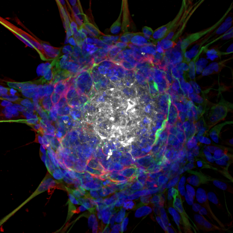
The project will also use surgical specimens provided through the collaboration with the hospital. The Łukasiewicz – PORT team will attempt to establish tumor cell cultures from these samples. This will allow the researchers to investigate which molecules are produced by the tumor cells and whether the same molecular signals can be detected in patients’ blood. Analyzing the surgical specimens will also provide a better understanding of how cancer cells interact with their surrounding microenvironment and why the immune system often fails to eliminate glioma effectively, despite the presence of immune cells within the tumor that are capable of attacking cancer.
This is particularly important because a tumor is not a uniform mass of cells. In addition to cancer cells, it contains what is known as the tumor microenvironment, which includes immune cells, blood vessel cells, fibroblasts, the extracellular matrix, and numerous signaling molecules. These components can both promote disease progression and reduce the effectiveness of treatment.
“Gliomas have a microenvironment that effectively suppresses the immune system. The tumor may contain cells capable of cytotoxic activity against cancer cells, but at the same time, components of the microenvironment trigger numerous mechanisms that inhibit this immune response,” says Dr. hab. Chodaczek.
The 4th Military Clinical Hospital in Wrocław will be responsible for patient recruitment and the clinical component of the project. As a non-interventional study, it will not involve any additional treatment-related procedures. Patients will follow the standard diagnostic pathway, including radiological imaging and histopathological examination. The only additional procedure performed for the purposes of the project will be a blood draw.
Artificial Intelligence and Precision Medicine
The data collected in the project will then be integrated and analyzed using bioinformatics tools and artificial intelligence methods. The goal is to develop an approach that more effectively combines different sources of diagnostic information, including radiological images, histopathological findings, and molecular data.
“Liquid biopsy is only one component. Equally important is whether the algorithms can integrate different diagnostic techniques into a single model and help determine the type and grade of the tumor—or whether a lesion detected through imaging is in fact cancerous,” explains Dr. hab. Chodaczek.
In addition to Dr. hab. Grzegorz Chodaczek’s team, the Łukasiewicz – PORT contribution to the project will involve the Genetic and Microbiological Diagnostics Laboratory, headed by Dr. Paweł Sega. The research aligns with the mission of the P4Health Center, which advances precision and personalized medicine.
Glioma Immunotherapy and Gamma Delta T Cells
In the previous project, Dr. hab. Chodaczek’s team sought to identify optimal donors of gamma delta T cells for potential use in glioma therapy. These cells are a distinct subset of immune cells. Unlike the more prevalent alpha beta T cells, which are typically associated with recognizing fragments of viruses or bacteria, gamma delta T cells can respond to signals indicating that something is wrong with a cell.
“They can be thought of as sensors of a cell’s metabolic state. Gamma delta T cells recognize when a cell begins to behave abnormally—for example, when it is under stress, undergoes metabolic changes, or becomes cancerous. This is precisely what makes them so interesting from the perspective of cancer immunology,” the researcher explains.
The researchers investigated why cells from some donors were more effective at destroying cancer cells than those from others. The project identified a set of proteins and genes that may determine how well a given T-cell culture performs under laboratory conditions.
The project has officially concluded, but the researchers are continuing their work. “We generated terabytes of data and are still analyzing it. This is not a process that can be completed within a year. We may identify a protein or gene that affects T-cell reactivity, but then we need to design further experiments and confirm whether it is truly significant. That is how new research questions emerge,” says Dr. hab. Chodaczek.
Even optimally prepared immune cells may prove ineffective if a patient’s tumor lacks the features that enable them to recognize it. As the researcher emphasizes, understanding the biology of tumors—their differences, the molecules they produce, and the mechanisms they use to evade immune surveillance—is therefore just as important as developing a potential therapy.
Research on T cells has shown that the effectiveness of a potential therapy depends not only on the properties of the immune cells but also on the individual characteristics of the tumor itself. The PROGLIO project will investigate whether characteristic signals released by gliomas can be detected in the blood and integrated with imaging findings and tumor tissue analysis. In the future, this approach could enable more accurate tumor assessment before treatment begins and support the selection of the most appropriate therapy.
“If we could identify a tumor-specific signature in the blood, that alone would be a significant achievement. If we could also develop an algorithm integrating radiological, histopathological, and molecular data, we would take an important step toward more accurate brain tumor diagnostics and, in the future, better treatment selection,” the researcher concludes.